Friday, September 25, 2015

Histology Staining Ch: Vocabulary

Microtome: a specifically designed slicing machine upon which samples are placed to be cut with a steel knife.

Autoradiography: process in which radioactively tagged precursrs of the molecule are incorporated by cells and tissue before fixation.

Eosin: a type of acid dye - which carries a net negative charge on its colored portion.

Basic Dye: carries a net positive charge on its colored portion.

Tuesday, September 15, 2015

Eyewire

I found something last weekend that has me excited. The kind of excited that has you talking about it, mulling over it, thinking about it when your suppose to be studying something else.

Let me tell you what it is. It is Eyewire - an opportunity for anyone to help advance neuroscience.

One of the most challenging aspects of neuroscience is understanding what the brain (or in this case - retina) -at the cellular level - looks like. Where are the synapses? Which neurons connect with which neurons? And so on. One of the ways to find out is to assemble 3D models or reconstructions of sections of the neural system.

The process of reconstructing every neuron & the synappses it forms with others & their supporting cells..........is lengthly and time consuming to say the least. One lab has come up with a way to speed up the process and allow anyone to contribute to groundbreaking science!

Eyewire: an online game where you can reconstruct neurons from electron micrographs! Its free, easy to learn, social (there's a live chat, a blog, a FB group, etc), has a beautiful and easy to use user interface, and the player is contributing to the one of the next great scientific  acheivements!

Find out for yourself, the link below is to the blog, from there you can also watch videos and visit the online game.
http://blog.eyewire.org/about/

Link to Histology: viewing thousands of cells in 3D! By building the 3D models, we will discover so many new things! Histology on a grand scale!
Friday, September 11, 2015

An Encounter with 3D Modeling of the Cortex

If you are like me, You prefer to experience things in 3D rather than in 2D. So when the opportunity comes to do so in the microscopic world of Biology, you get excited. Especially, when its a highly detailed, informative, complex, intricate, colorful, moving, beautiful section of the prefrontal cortex. Thats right, someone took the time to reveal such a beautiful piece of art. As art alone it is beautiful & complex, but when you think about what goes on in this microscopic part of the brain and how it interacts with the rest of the brain, and what the results of those interactions are?.................. Awesome!

If you would like to see the video this is from here is the link: https://youtu.be/8YM7-Od9Wr8


The 3D model was originally a part of the following paper:
"Saturated Reconstruction of a Volume of Neocortex" by: Narayanan Kasthuri8correspondenceemail, Kenneth Jeffrey Hayworth9, Daniel Raimund Berger, Richard Lee Schalek, José Angel Conchello, Seymour Knowles-Barley, Dongil Lee, Amelio Vázquez-Reina, Verena Kaynig, Thouis Raymond Jones, Mike Roberts10, Josh Lyskowski Morgan, Juan Carlos Tapia11, H. Sebastian Seung12, William Gray Roncal13, Joshua Tzvi Vogelstein14, Randal Burns, Daniel Lewis Sussman, Carey Eldin Priebe, Hanspeter Pfister, Jeff William Lichtmancorrespondenceemail

Friday, September 4, 2015

Why I picked "Art...On a Microscopic Scale" as the Title of this Blog

Histology is the study of the tissues that make up organisms and the cells that make up those tissues. Since we rely heavily on vision as humans, this means Histology involves a lot of visual data. 


  • Cross sections of tissue mounted on slides
  • 3D reconstructions of various structures
  • 3D intact structures that have been made transparent to an extent and per-fused with a particular marker
  • Images of tissues

The reason I am most looking forward to this class, is simply the beauty and artistry that I will get to see. Yes, the information I will learn is interesting and I am eager to learn it. But there is a quality to histology that is even more attractive to me - art. The beauty with which each structure is created is unique and similar to many styles of art. 


Renal Medulla - 1
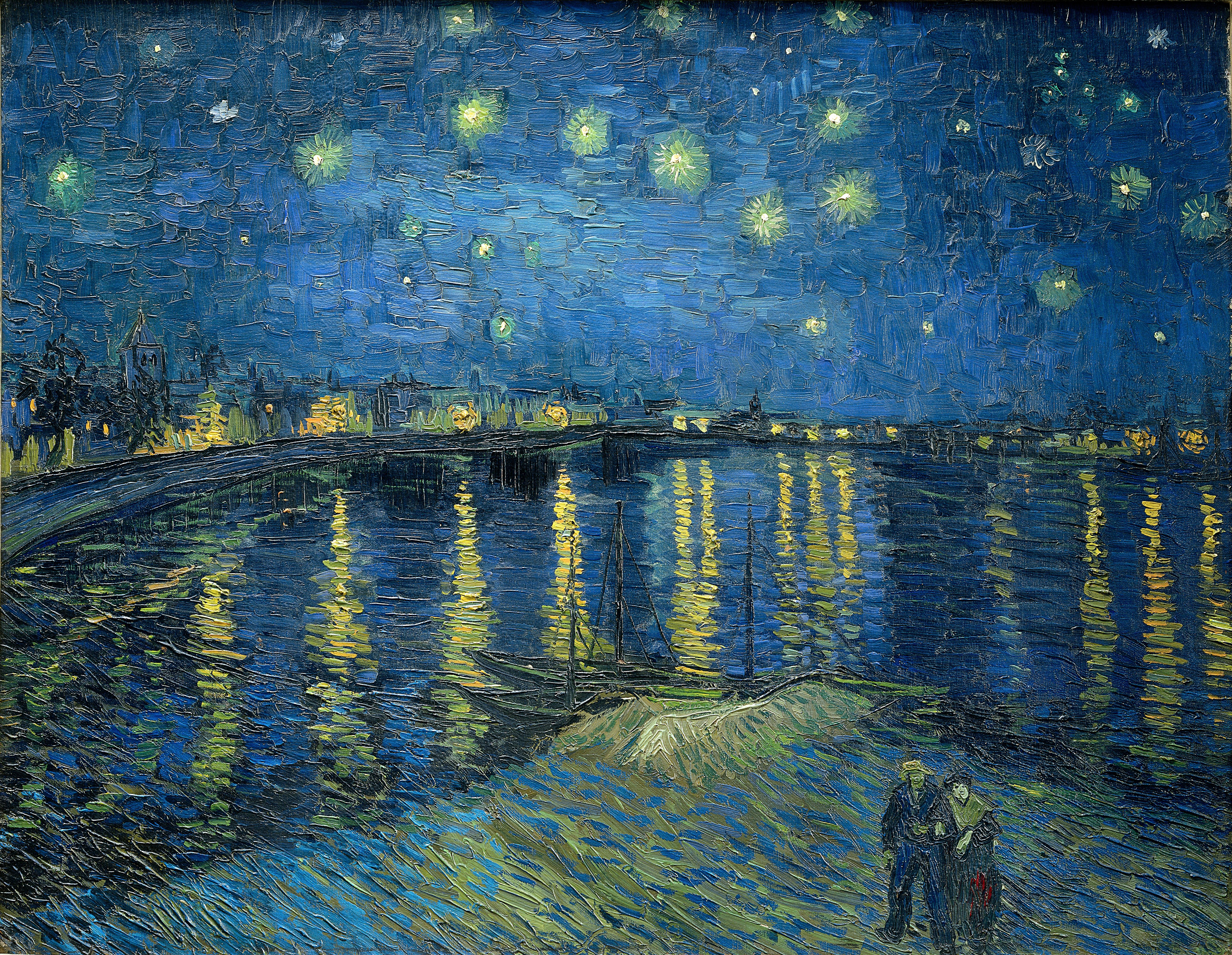
Its a long jump, but the pattern I see here in the renal medulla reminds me of things like Van Gogh's Starry Night over the Rhone, Aboriginal Dreamtime art, and the painting style of Monet. An organic, abstract pattern of various sizes of dots. 


Camille Monet and a Child in the Artist' Garden in Argenteuil - 4
Starry Night over the Rhone - 5
Dreamtime Sisters - 6


Another Reason I appreciate Histology as a type of art - is because the slides themselves have qualities that, if described with terms typically applied to art, make them a type of art. One of my favorite TV shows is NCIS.  A character on the show (Abbey) has decorated her office with images taken with different lab techniques. The images themselves are visual data that she had used to solve cases in the past, but they are also beautiful in their own right. Similarly - slides of tissue can be as well.


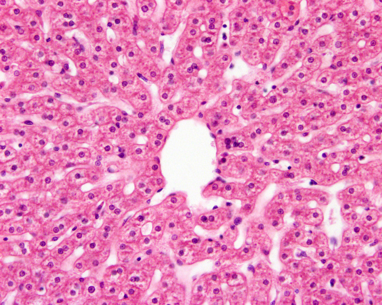
Central Vein of Liver - 2
The white space (the cavity within the central vein) has abstract lines  radiating out of it, like a sun or the petals of a flower. The deeper purple of the nuclei add another level of design to the image. the image is busy, but the blank space in the middle allows the eye to rest - this could be a good focal piece. 


Brain (Cerebellum) - 3
This is a beautiful design you might incorporate into a choral reef themed room, hang on the wall, design a modern coat hanger based off of. The design is in different shades of the same color. If this were a painting, I would say the darker purple was used to create an outline of the design of he lighter purple. Or maybe the dark purple creates a design of the interlocking and overlapping leaves of a tropical rain forest. The white spaces would be the light shining through the leaves. 

The body is a complex thing of machinery, a powerful computer, an organism. These are all ideas that have been used to describe the body in the past and influence the way we think about the body. Whether it be human, mamal, reptile, or mullusc. I would like to present another lense through which to view cells, tissues, organs, and organisms as a whole. As beautiful pieces of art. Because they are complex and beautiful, no one can deny that. 


References: 

  1. http://www.siumed.edu/~dking2/crr/RN021b.htm
  2. https://embryology.med.unsw.edu.au/embryology/index.php?title=File:Liver_histology_101.jpg
  3. http://www.sporcle.com/games/sbme/histology-slideshow
  4. http://www.wikiart.org/en/claude-monet/madame-monet-and-child
  5. https://en.wikipedia.org/wiki/The_Starry_Night
  6. http://www.aboriginalartstore.com.au/artists/colleen-wallace-nungari/dreamtime-sisters-191/