Friday, October 30, 2015

Lunar Histology

So Im on a camping trip with my pathfinder club, its Friday night & we are setting up camp in the dark. Im in camping mode - school mode has long been forgotton, and I am enjoying the break. Its a beautiful night, the sky is clear, the air is cold & refreshing, and the air is so much cleaner. The moon is almost full & is extra bright. I stop to admire the moon, in all of its bright clarity. Its a beautiful moon, and what does my mind come up with? "Oh, its a neutrophil!" (I genuinly ment it, no prior thought)

Then I realized what utter geekness had just come out of my mouth! It was hilarious, I thought I had switched modes, but I guess my brain was still filled with the facts for the exam that morning. Anyway, it was hilarious. So I found a few people around camp who could appriciate the humor.

I also texted a friend who is in medical school. After confirming how geeky my comment had been, and thoroughly rubbing it in my face, she commented that the moon actually is a lymphocyte most of the time. One of the staff camping with us, commented that sometimes the moon is sickled.

Oh what a bunch of geeks we are! It makes life so much more hilarious! Studying biology is fun, interesting, and a great challenge.........the effects it has on my brain are hilarious sometimes.

Friday, October 23, 2015

Design - Inspired by Histology

This post is a light hearted one to emphasize histology through the artistic lens. Take a break from studying, understanding, memorizing, identifying.......take a step back......and wonder at the abstract beauty of Histology. Some of the pictures are of actual tissue, some are histology inspired (whether for asthetic or humor), and some are a little of both. Enjoy. 

1

2
3
4
5


6


7


8


References: 

  1. http://www.anatomybox.com/wp-content/uploads/2012/12/Histology-Plates1.jpg
  2. http://www.ultralighthistology.com/images/colondetail4.jpg
  3. https://blogger.googleusercontent.com/img/b/R29vZ2xl/AVvXsEgrOkCXtYUMpLHsJVq72DGRXoY7TC_xm6RGtOH2rWRS2711tqXnwt6rATL-K4EnrW-PhLkX0o8ecaI1YYPyIRVg6BsZrk2ynsxCdGWatDtlOyqyoTfpfLAaXVZ3OvSBIDUuuuiAonS2WfU/s1600/4-histology-nail-art.jpg
  4. https://pbs.twimg.com/media/CBRhgt-UsAAAvQ5.jpg
  5. http://41.media.tumblr.com/98f13856b33508413329a572482f6306/tumblr_mg5tmyUyit1s24chqo1_1280.jpg
  6. http://farm1.static.flickr.com/62/186818508_ceb68c9987.jpg
  7. https://s-media-cache-ak0.pinimg.com/originals/98/b2/d0/98b2d0ce04bf3ab7847a78faa8890888.jpg
  8. http://www.histology-world.com/image/boneart2t.jpg

Friday, October 16, 2015

Histology Vocabulary

Schmidt-Lanterman cleft: the space between successive lamellae of the myelin.

Internodal segment: myelin between sequential nodes of Ranvier.

Neuregulin (Ngr1): growth factor that regulates myelin sheath thickness.

Transcription factor Sox -10: causes neural crest cells to develop into Schwann cells.

Tracts: the organizational label given to many axons going to or comming from a specific location - these are bundled together  and labeled a tract.

Neuropil: the meshwork of axons, sendrites, and glial processes associated with white matter.

Anterograde (Wallerian) degeneration: axon degeneration  distal to the site of injury.

Friday, October 9, 2015

Histology Reflection

Its interesting. Before this histology course, I was not sure what I was looking at when I looked at histology pictures online. I knew a few things here & there, but always doubted if I was correct. Now, after only a few Histology labs I understand what I am looking at. I am also more confident when I describe the structures I see through the lense of the microscope.  I am not saying that I can correctly  identify every structure in every slide that comes my way. What I am saying is, the practice of looking through the microscope at a variety of tissue samples has trained me to recognize patterns, similar structures, key differences, and other hints I can use to better understand what I am looking at.

I have enjoyed the Histology lab a lot more than I thought I would. Mostly, I was expecting the lab to be similar to the Plant Diversity lab I had experienced. A lab full of interesting things to look at and try to  draw......with not near enough time to see all of them in.  The perfect recipe for frustration. However this lab has far fewer samples required too look at per lab  session, and I have switched from drawing to taking photos of what I see through the microscope.  This provides me with the time to really look at each sample. To enjoy their beauty, appriciate their complexity, see the small details, ask questions & think of answers to those questions. I learn so much more this way, and I have time to enjoy the learning experience.

I am excited to gain more histology skills, so that I can incorporate them into my understanding of biology.

Sunday, October 4, 2015

The importance of Art to Science - an Encounter

Visual data & artistic representations help us better understand Biology and remind us that life is complex and beautiful!

  "Huntington disease is a progressive brain disorder that causes uncontrolled movements, emotional problems, and loss of thinking ability (cognition) (1)."
https://instagram.com/p/8MPtIiQBB9/

Mutations in the HTT gene that codes for the protein Huntingtin cause Huntington disease. The function of the protein is still unknown. The mutation is a CAG triplet repeat; normally this pattern is repeated 10 to 35 times within the gene, in individuals with huntington disease the CAG triplet is repeated 36 to 120 times within the gene. Individuals with the CAG triplet repeated between 36 and 39 times may or may not develop Huntington disease. The longer gene causes production of abnormally long Huntingtin proteins that are then "cut into smaller, toxic fragments that bind together and accumulate in neurons, disrupting the normal functions of these cells"(1).

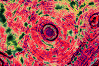
"This illustration shows potassium levels in astrocytes -- cells in your nervous system -- disrupted by Huntington’s Disease, a genetic disorder that causes the breakdown of nerve cells in the brain. In this artistic rendering, created by @TEDFellow Janet Iwasa and scientist Baljit Khakh, potassium is represented by glowing points of light. Those points show how potassium accumulates in astrocytes and not in the surrounding neuropil, contradicting the idea that Huntington’s Disease is caused by neuronal dysfunction. To see more of Janet’s scientific illustrations, visit go.ted.com/huntingtons" - TED original Instram Commemt (2). 


Originally this was what my topic was going to be for this blog, then I visited the TED link in the Instagram post. I watched a video about animating biological processes. This caught my attention aswell. The presenter had an amazing idea (as most TED speakers do) to create open source, simple to use, animation software that would allow biology researchers the ability to animate their hypothesis. This is increadible, it would allow researches to see their ideas and/or results from a totally new perspective; help students better understand complex and interconnected biological processes; shed light on possibly incorrect hypotheses (like the photo above does); and reveal the true beauty of biology at the molecular level. The software is called Molecular Flipbook, and is free to download.

Then I thought about the beautiful artwork on my Cell & Molecular Biology book. The artwork is that of David Goodsell - an associate professor in the Department of Molecular Biology at the Scripps Research Institute in La Jolla, California. It is so important to have people creating visual representations of the cellular processes. Visuals help us better understand what is going on. Visuals are also very beautiful. 

Don't think of Biology as a bunch of facts to memorize, medical information, strange protein names, or random multi-cellular critters. There is a huge potential for art inherent in the study of Biology. This is because, living things are complex and beautiful. Take advantage of all the art linked to biology, enjoy the art, enjoy the beauty of life. 


References: 
  1. http://ghr.nlm.nih.gov/condition/huntington-disease
  2. https://instagram.com/p/8MPtIiQBB9/
  3. http://www.ted.com/talks/janet_iwasa_how_animations_can_help_scientists_test_a_hypothesis?utm_source=instagram&utm_medium=referral&utm_campaign=IGTalks
  4. https://www.molecularflipbook.org/
  5. http://mgl.scripps.edu/people/goodsell/
  6. https://en.wikipedia.org/wiki/David_Goodsell

Friday, September 25, 2015

Histology Staining Ch: Vocabulary

Microtome: a specifically designed slicing machine upon which samples are placed to be cut with a steel knife.

Autoradiography: process in which radioactively tagged precursrs of the molecule are incorporated by cells and tissue before fixation.

Eosin: a type of acid dye - which carries a net negative charge on its colored portion.

Basic Dye: carries a net positive charge on its colored portion.

Tuesday, September 15, 2015

Eyewire

I found something last weekend that has me excited. The kind of excited that has you talking about it, mulling over it, thinking about it when your suppose to be studying something else.

Let me tell you what it is. It is Eyewire - an opportunity for anyone to help advance neuroscience.

One of the most challenging aspects of neuroscience is understanding what the brain (or in this case - retina) -at the cellular level - looks like. Where are the synapses? Which neurons connect with which neurons? And so on. One of the ways to find out is to assemble 3D models or reconstructions of sections of the neural system.

The process of reconstructing every neuron & the synappses it forms with others & their supporting cells..........is lengthly and time consuming to say the least. One lab has come up with a way to speed up the process and allow anyone to contribute to groundbreaking science!

Eyewire: an online game where you can reconstruct neurons from electron micrographs! Its free, easy to learn, social (there's a live chat, a blog, a FB group, etc), has a beautiful and easy to use user interface, and the player is contributing to the one of the next great scientific  acheivements!

Find out for yourself, the link below is to the blog, from there you can also watch videos and visit the online game.
http://blog.eyewire.org/about/

Link to Histology: viewing thousands of cells in 3D! By building the 3D models, we will discover so many new things! Histology on a grand scale!