


Alveolar ducts - elongated airways that have almost no ducts, only alveoli as their peripheral boundary.
Alveolar Sacs - spaces surrounded by clusters of alveoli.
Ciliated cells - tall columnar cells with cilia that project into the mucus covering the surface of the epithelium
Goblet cells - synthesize and secrete mucus
Brush cells - a general name for cells in the respiratory tract that bear short, blunt microvilli
Small granules cells - resemble basal cells but contain secretory granules. These are endocrine cells of the diffuse neuroendocrine system
Basal cells - stem cells from which the other cell types
Olfactory receptor cells - are bipolar olfactory neurons that span the thickness of the epithelium and enter the central nervous system
Supporting or sustentacular cells - columnar cells that are similar to neuroglia cells and provide mechanical and metabolic support to the olfactory receptor cells. They synthesize and secrete odorant binding.
While researching for one of my Histology projects, I stumbled upon this great resource.
http://www.pathpedia.com
This is a global database for pathophysiology, currated by pathologists! It has wonderful collections of histology slides, with very informative captions. It also has an immunology database that I am eager to explore further. The site is for Doctors, researchers, laboratory technicians, medical students, and biology students. So the site carries indepth information and often either has peer reviewed articles or links to them. The site is also open to the public, free of charge, however it is geared towards those who study biology.
Finally, I have found a site that has quality pictures and information I can use for my histology projects. I also will be spending much more time further exploring the website. Hopefully, Ill restrain myself from doing so.....till after I finish my projects!
I hope this post introduces this amazing resource to someone else looking for credible histology slides & information.
Esophagus: the esophagus is a fixed muscular tube that delivers food and liquid from the pharynx to the stomach
Submucosa: the submucosa consists of a dense irregular connective tissue layer containing blood and lymphatic vessels, a nerve plexus, and occasional glands
Muscularis externa: in most parts of the digestive tract the muscularis externa consists of two concentric and relatively thick layers of smooth muscle
Mucosa: the mucosa consists of a lining epithelium, an underlying connective tissue called the lamina propria and the muscularis mucosa composed of smooth muscle
Serosa: the serosa is a serous membrane consisting of a simple squamous epithelium the mesothelium and a small amount of underlying connective tissue
Gastric Mucosa: longitudinal submucosal folds, rugae, allow the stomach to distend when filled
The busy, over-streached, suffering the snow ball effect, biology student's Lament
Histology: Epithelium, Basal Lamina, Tunica Adventita, Neuromuscular Junction, Eosinophile, Cadherin, Zonula Occludens, Lymph node, Circum Vitallea, hair follicle
"Wow, this is so interesting, I cant wait to study it, and find out as much as I can. Im gonna be able to fill in so many of the gaps in my knowledge. This is going to be fun! Im gonna read, take notes, make colorful note pages, watch histology vides, look at awesome slides of what Ive been wondering about, ask Questions, watch TED TALKs on Histology (always amazing), draw cool designs influenced by histology, hold discussions with my peers about histology, use the research projects to journey down the rabbit hole, and read other chapters in my text book - just cause a picture caught my eye!"
Night A:
"Finally done with my classes for the day! Now I can read my histo...."Hold on, you have an assignment due for this other class tonight, and a quiz, and that Discussion Post, oh dont forget to do what you said you would do for that particular person.
"Ugh, finally done....now I can read my histology book!" (Ten minutes later) the biology student is peacefully dreaming of epithelial linings, cilia, free surfaces & and basal lamina......with her head nestled in her histology text book.
Night B:
"Im so tired, I havent had a break all day, I dont remember eating lunch. I just want to sleeeeeeeep!" A friendly advise giver "sleep is important, you need to be rested, go to bed." "No, I have so many assignments I have to do!" Proceeds to try to work on the computer, zones out, watches videos, tries to read & has to read the same paragraph three times, contemplates going to bed.....re-musters up focus, has a productive 30 minutes, then.............. "what? Its 3am? It was 1:30 two seconds ago!" Defeated, stumbles to bed.
Night C:
"I cant take it anymore! Im going to study Histology!" Starts reading histology book (or slides), enjoys reading the new information, begins to get into the text - memorizing, developing questions, associating new info with old info......
"You should do this project!" "You forgot to that lab report" "You havent excersized in a week" "You need to write that paper!"
Student is now feeling guilty for studying Histology instead of completing all the other work. Puts book aside & tries to work on a paper....proceeds very slowly, because mind is still on the histology studies.
Oh Histology, to study you more, to study you more.
(The author of this rambling post is not looking for special favors, sympathy, or empathy. The author only hopes someone will be entertained by this post.)
So Im on a camping trip with my pathfinder club, its Friday night & we are setting up camp in the dark. Im in camping mode - school mode has long been forgotton, and I am enjoying the break. Its a beautiful night, the sky is clear, the air is cold & refreshing, and the air is so much cleaner. The moon is almost full & is extra bright. I stop to admire the moon, in all of its bright clarity. Its a beautiful moon, and what does my mind come up with? "Oh, its a neutrophil!" (I genuinly ment it, no prior thought)
Then I realized what utter geekness had just come out of my mouth! It was hilarious, I thought I had switched modes, but I guess my brain was still filled with the facts for the exam that morning. Anyway, it was hilarious. So I found a few people around camp who could appriciate the humor.
I also texted a friend who is in medical school. After confirming how geeky my comment had been, and thoroughly rubbing it in my face, she commented that the moon actually is a lymphocyte most of the time. One of the staff camping with us, commented that sometimes the moon is sickled.
Oh what a bunch of geeks we are! It makes life so much more hilarious! Studying biology is fun, interesting, and a great challenge.........the effects it has on my brain are hilarious sometimes.
 |
| 1 |
 |
| 2 |
 |
| 3 |
 |
| 4 |
 |
| 5 |
 |
| 6 |
 |
| 7 |
 |
| 8 |
Schmidt-Lanterman cleft: the space between successive lamellae of the myelin.
Internodal segment: myelin between sequential nodes of Ranvier.
Neuregulin (Ngr1): growth factor that regulates myelin sheath thickness.
Transcription factor Sox -10: causes neural crest cells to develop into Schwann cells.
Tracts: the organizational label given to many axons going to or comming from a specific location - these are bundled together and labeled a tract.
Neuropil: the meshwork of axons, sendrites, and glial processes associated with white matter.
Anterograde (Wallerian) degeneration: axon degeneration distal to the site of injury.
Its interesting. Before this histology course, I was not sure what I was looking at when I looked at histology pictures online. I knew a few things here & there, but always doubted if I was correct. Now, after only a few Histology labs I understand what I am looking at. I am also more confident when I describe the structures I see through the lense of the microscope. I am not saying that I can correctly identify every structure in every slide that comes my way. What I am saying is, the practice of looking through the microscope at a variety of tissue samples has trained me to recognize patterns, similar structures, key differences, and other hints I can use to better understand what I am looking at.
I have enjoyed the Histology lab a lot more than I thought I would. Mostly, I was expecting the lab to be similar to the Plant Diversity lab I had experienced. A lab full of interesting things to look at and try to draw......with not near enough time to see all of them in. The perfect recipe for frustration. However this lab has far fewer samples required too look at per lab session, and I have switched from drawing to taking photos of what I see through the microscope. This provides me with the time to really look at each sample. To enjoy their beauty, appriciate their complexity, see the small details, ask questions & think of answers to those questions. I learn so much more this way, and I have time to enjoy the learning experience.
I am excited to gain more histology skills, so that I can incorporate them into my understanding of biology.
 |
| https://instagram.com/p/8MPtIiQBB9/ |
Microtome: a specifically designed slicing machine upon which samples are placed to be cut with a steel knife.
Autoradiography: process in which radioactively tagged precursrs of the molecule are incorporated by cells and tissue before fixation.
Eosin: a type of acid dye - which carries a net negative charge on its colored portion.
Basic Dye: carries a net positive charge on its colored portion.
I found something last weekend that has me excited. The kind of excited that has you talking about it, mulling over it, thinking about it when your suppose to be studying something else.
Let me tell you what it is. It is Eyewire - an opportunity for anyone to help advance neuroscience.
One of the most challenging aspects of neuroscience is understanding what the brain (or in this case - retina) -at the cellular level - looks like. Where are the synapses? Which neurons connect with which neurons? And so on. One of the ways to find out is to assemble 3D models or reconstructions of sections of the neural system.
The process of reconstructing every neuron & the synappses it forms with others & their supporting cells..........is lengthly and time consuming to say the least. One lab has come up with a way to speed up the process and allow anyone to contribute to groundbreaking science!
Eyewire: an online game where you can reconstruct neurons from electron micrographs! Its free, easy to learn, social (there's a live chat, a blog, a FB group, etc), has a beautiful and easy to use user interface, and the player is contributing to the one of the next great scientific acheivements!
Find out for yourself, the link below is to the blog, from there you can also watch videos and visit the online game.
http://blog.eyewire.org/about/
Link to Histology: viewing thousands of cells in 3D! By building the 3D models, we will discover so many new things! Histology on a grand scale!
 |
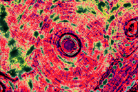
| Renal Medulla - 1 |
 |
| Camille Monet and a Child in the Artist' Garden in Argenteuil - 4 |
 |
| Starry Night over the Rhone - 5 |
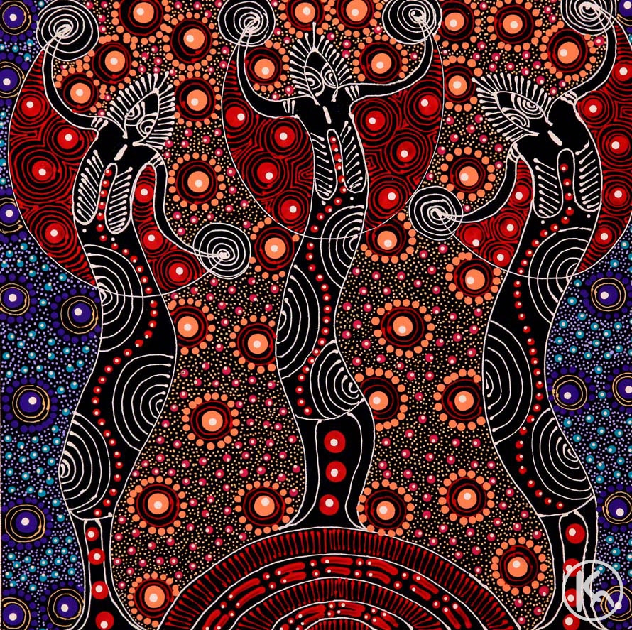 |
| Dreamtime Sisters - 6 |
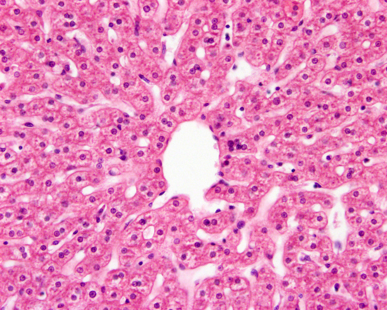 |
| Central Vein of Liver - 2 |
 |
| Brain (Cerebellum) - 3 |